Potato Salad With Raisins - Raisins In Potato Salad : Kale & Roasted Sweet Potato ... / Add a sprinkling of salt and lemon juice to the water as it boils so that the. . Stir lightly with a rubber spatula until evenly coated with dressing. Последние твиты от potato salad with raisins (@ianbruh76). ½ cup of cooked quinoa (about 90g). Enjoy this easy carrot salad with raisins along with a family meal or holiday dinner. I sometimes put raisins in my potato salad but more often i will add white grapes cut in half or craisins for that little sweet, along with almonds or walnuts or pecans. Potato salad with raisin sounds like the sort of thing people in that shitstain state of ohio would eat. with raisins, it's the chewiness and somewhat gritty texture mixed with the carby softness of potatoes that i find revolting. This twist on potato salad is packed with flavor from sweet raisins, fresh mint, and tangy yogurt. It is an easy dish with an simmer until potatoes are ...
Dapatkan link
Facebook
X
Pinterest
Email
Aplikasi Lainnya
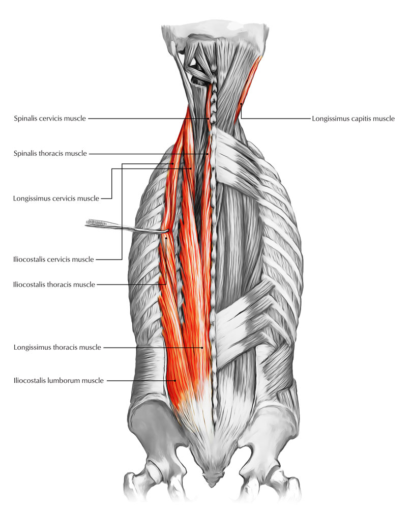
Back Muscles Anatomy - Anatomy Of Back Muscles Diagram - The muscles of the back can be arranged into 3 categories based on their location:
Dapatkan link
Facebook
X
Pinterest
Email
Aplikasi Lainnya
Back Muscles Anatomy - Anatomy Of Back Muscles Diagram - The muscles of the back can be arranged into 3 categories based on their location:. These muscles include the large paired muscles in the lower back, called erector spinae, which help hold up the spine, and gluteal muscles. The vertebral column consists of 33 vertebrae which can be split up into 5 continuous sections. Anatomy of the back muscles. To control the posture of the entire body. We think this is the most useful anatomy picture that you need.
Back muscles, functions and exercises: The muscles of the back can be arranged into 3 categories based on their location: Anatomy of the back muscles. Anatomynote.com found anatomy of back muscles diagram from plenty of anatomical pictures on the internet. On this page, youll learn about each of these muscles, their locations, and functional anatomy.
Back Muscles 28 Major Muscles Of The Back Earth S Lab from www.earthslab.com The back anatomy includes the latissimus dorsi, trapezius, erector spinae, rhomboid, and the teres significant. Related posts of back muscles chart muscle anatomy for artists. Elbow muscle anatomy mri 12 photos of the elbow muscle anatomy mri elbow muscle anatomy axial, elbow muscle anatomy mri, human muscles, elbow muscle anatomy axial, elbow muscle anatomy mri. We hope this picture anatomy of back muscles diagram can help you study and research. The intrinsic back muscles are found deeper to the extrinsic muscles, separated from them by the thoracolumbar fascia. Anatomy of the back muscles the latissimus dorsi muscles (also known as the lats) are the largest muscles of the back. They provide movements of the spine , stability to the trunk, as well as the coordination between the movements of the limbs and trunk. Back muscles are divided into two specific groups:
Since the all the back muscles originate in embryo (fetus) form by locations other than the back, muscles in the superficial, as well as, intermediate groups, are extrinsic muscles.
The semispinalis dorsi and semispinalis capitis muscles also extend the back. The extrinsic muscles superficial extrinsic muscles connect your upper … This blog post article is an overview of the muscles of the lumbar spine of the trunk. They provide movements of the spine , stability to the trunk, as well as the coordination between the movements of the limbs and trunk. Lower back muscle anatomy includes the multifidus, longissimus, spinalis, and quadratus lumborum. The back anatomy includes the latissimus dorsi, trapezius, erector spinae, rhomboid, and the teres significant. The vertebral column consists of 33 vertebrae which can be split up into 5 continuous sections. These muscles include the large paired muscles in the lower back, called erector spinae, which help hold up the spine, and gluteal muscles. Superficial back muscles, intermediate back muscles and intrinsic back muscles.the intrinsic muscles are named as such because their embryological development begins in the back, oppose to the superficial and intermediate back muscles which develop elsewhere and are therefore classed as extrinsic muscles. Muscle anatomy for artists 12 photos of the muscle anatomy for artists female muscle anatomy for artists, human muscle anatomy for artists, male muscle anatomy for artists, muscle anatomy for artists, human muscles, female muscle anatomy for artists, human muscle anatomy for. The pelvic floor muscles also help increase this pressure, which provides stability to the spine and trunk. The latissimus dorsi muscles (also known as the lats) are the largest muscles of the back. We hope this picture anatomy of back muscles diagram can help you study and research.
Related posts of back muscles chart muscle anatomy for artists. Anatomy of the back muscles the latissimus dorsi muscles (also known as the lats) are the largest muscles of the back. These muscles give height and breadth to back development. The erector spinae muscles, for example, extend the back (bend it backward) and side bend the back. On this page, you'll learn about each of these muscles, their locations and functional anatomy.
Human Anatomy Of Muscles Detailed Diagram Of Back Muscles Royalty Free Cliparts Vectors And Stock Illustration Image 13475146 from previews.123rf.com The lumbar region of the spine, more commonly known as the lower back, is situated between the thoracic, or chest, region of the spine, and the sacrum. Three types of back muscles that help the spine function are extensors, flexors and obliques. Anatomynote.com found anatomy of back muscles diagram from plenty of anatomical pictures on the internet. Leaning back to straight vertical and all points in between. Related posts of back muscles chart muscle anatomy for artists. These are the muscles that are farther from the surface, closer to the internal organs and the spine. The human back, also called the dorsum, is the large posterior area of the human body, rising from the top of the buttocks to the back of the neck. The latissimus dorsi muscles (also known as the lats) are the largest muscles of the back.
As a general group, they extend from the neck to the sacrum and fulfill a basic and fundamental function:
(2017, elsevier) should be consulted. All about the back muscles the back anatomy includes the latissimus dorsi, trapezius, erector spinae, rhomboid, and the teres major. Several small muscles in the cervical area of the vertebral column are also important. The back consists of the spine, spinal cord, muscles, ligaments, and nerves. Related posts of muscles of the lower back and buttocks diagram elbow muscle anatomy mri. It is the surface of the body opposite from the chest.the vertebral column runs the length of the back and creates a central area of recession. Back muscles anatomy the surface muscles of the upper back include the trapezius muscles (traps) and posterior deltoids. Lumbar spine anatomy video understanding the anatomy of your lower spine can help you communicate more effectively with the medical professionals who treat your lower back pain. Anatomy of the back muscles. The anatomy of the back refers to the muscles of the back, as well as the bones of the scapulae, ribcage, and spine.covering an expanse from the neck to the tailbone, the back muscles are responsible for a broad range of functions, from extending the spine to shrugging the shoulders.these muscles facilitate movement by attaching to one or more bones of the back, either to the spinous processes. Elbow muscle anatomy mri 12 photos of the elbow muscle anatomy mri elbow muscle anatomy axial, elbow muscle anatomy mri, human muscles, elbow muscle anatomy axial, elbow muscle anatomy mri. The back muscles are anatomically layered into superficial (extrinsic) and deep (intrinsic) muscles. Back muscles are divided into two specific groups:
To control the posture of the entire body. The erector spinae muscles, for example, extend the back (bend it backward) and side bend the back. The small muscles of the vertebrae (the multifidi and rotators) help rotate, extend, and side bend the back. As a general group, they extend from the neck to the sacrum and fulfill a basic and fundamental function: The muscles of the back can be arranged into 3 categories based on their location:
The Back Muscle Anatomy Human Anatomy from 2.bp.blogspot.com Superficial back muscles, intermediate back muscles and intrinsic back muscles.the intrinsic muscles are named as such because their embryological development begins in the back, oppose to the superficial and intermediate back muscles which develop elsewhere and are therefore classed as extrinsic muscles. We think this is the most useful anatomy picture that you need. We hope this picture anatomy of back muscles diagram can help you study and research. Anterior rami of spinal nerve innervate them. The lumbar region of the spine, more commonly known as the lower back, is situated between the thoracic, or chest, region of the spine, and the sacrum. This blog post article is an overview of the muscles of the lumbar spine of the trunk. Three types of back muscles that help the spine function are extensors, flexors and obliques. On this page, you'll learn about each of these muscles, their locations and functional anatomy.
The lumbar region of the spine, more commonly known as the lower back, is situated between the thoracic, or chest, region of the spine, and the sacrum.
The human back, also called the dorsum, is the large posterior area of the human body, rising from the top of the buttocks to the back of the neck. The anatomy of the back refers to the muscles of the back, as well as the bones of the scapulae, ribcage, and spine.covering an expanse from the neck to the tailbone, the back muscles are responsible for a broad range of functions, from extending the spine to shrugging the shoulders.these muscles facilitate movement by attaching to one or more bones of the back, either to the spinous processes. To control the posture of the entire body. The breadth of the back is created by the shoulders at the top and the pelvis at the bottom. It is the surface of the body opposite from the chest.the vertebral column runs the length of the back and creates a central area of recession. Anatomy of the back muscles. The back muscles are divided into two large groups: The muscles of the back can be arranged into 3 categories based on their location: Muscles of the lumbar spine. The extrinsic muscles that are associated with upper extremity and shoulder movement, and the intrinsic muscles that deal with movements of the vertebral column. We hope this picture anatomy of back muscles diagram can help you study and research. Anatomynote.com found anatomy of back muscles diagram from plenty of anatomical pictures on the internet. Back muscles anatomy the surface muscles of the upper back include the trapezius muscles (traps) and posterior deltoids.


Komentar
Posting Komentar